
Silva Lab

Reprogramming cells to unlock developmental potential with embryo models
Embryo models will generate high-quality cells for medical applications
For many years, our lab has focused on understanding the biology of nuclear reprogramming, with the ultimate goal of using generated induced pluripotent stem cells (iPSCs) for medical applications. Despite significant efforts by labs worldwide, approaches like pluripotent stem cell differentiation, direct/forward programming, and cell transdifferentiation have not yielded cell products with the desired molecular identities and physiological properties. We understand that during development, cells establish stage-specific epigenetic signatures that influence transcriptional profiles and phenotypes. These are influenced by the specific context which include the precise signalling cues present during development, which cannot be replicated by traditional pluripotent stem cell differentiation methods.
Our studies on nuclear reprogramming and subsequently on applying this to embryo modelling are showing a new path as to how this limitation might be overcome. In 2019 (Stuart et al.), we showed that STAT3-mediated reprogramming to induced pluripotency proceeds through transient intermediates that molecularly and functionally resemble early embryonic cells preceding conventional pluripotency. Although this was a side finding in the indicated study, it was an “aha” moment, as it revealed that reprogramming could provide access to cells with enhanced developmental competence. This may also have parallels to natural embryonic development.
From embryo founder-like cells to Advanced Embryo Models

Video showing reprogramming to naive pluripotency using activated STAT3. Reprogramming proceeds through transient intermediates resembling early embryonic cells, marked by transient Gata6-driven GFP expression (Stuart et al 2019). Neither the starting cells nor the resulting iPSCs express Gata6; expression is restricted to the reprogramming intermediates.
Pioneering work by Magdalena Zernicka-Goetz and Alfonso Martinez-Arias labs showed that pluripotent stem cells can self-organise and model aspects of embryonic development. Building on this concept and our expertise and insights in nuclear reprogramming, we hypothesised that using our reprogrammed cells, which exhibit molecular and phenotypic features of early embryonic cells, as the starting population would provide greater developmental competence for self-organisation, enabling efficient and faithful modelling of embryonic development.
We therefore set to reprogram mouse and human pluripotent stem cells, including embryonic stem cells (ESCs) and iPSCs, into what we termed “induced embryo founder-like cells (iEFCs)” (Li et al, 2023; Guo et al, 2024; Li et al, 2024; Li et al 2025; Chen et al, 2025). These cells resemble earlier stages of embryo development compared to pluripotent stem cells and express combinations of antagonistic lineage-associated transcription factors, including NANOG, GATA6, CDX2 and OCT4. They also acquire competence to specify all blastocyst fates; trophectoderm/trophoblast, primitive endoderm/hypoblast and naïve epiblast.

Generation of iEFCs, marked by activation of early embryo markers Gata6 (white) and Cdx2 (red). Sox2 expression is shown in green (Li et al, 2024; Li et al 2025).
When let alone in suspension, these cells (iEFCs) were found to aggregate and self-organise into embryo-like structures that were found to undergo coordinated developmental progression, closely modelling key features of embryogenesis.

iEFCs aggregate and self-organize into a high-fidelity embryo model (Li et al 2025)
We have also extended this approach to human embryo modelling. In our recent study (Chen et al., 2025), STAT3-reprogrammed human cells self-organised into embryo-like structures that modelled key features of early post-implantation human embryonic development. We now want to harness the potential of our embryo models as platforms for generating high-quality cells to open new opportunities to advance medicine.
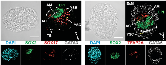
Carnegie Stage 5 (CS5)-like structures illustrating the capacity of our system to model early stages of human post-implantation development
Enjoy the videos below of our mouse embryo model!
The first two videos capture the intact structures of our complete embryo model, comprising the placenta, yolk sac, and embryo. The third video shows the embryo model with the yolk sac and placenta removed, exposing the embryo proper, now enclosed only by the amnion.


Click below to view lab publications


Sponsors :
The background image shows beautiful Guangzhou at night